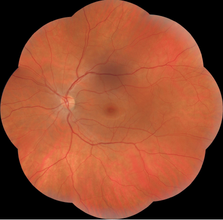
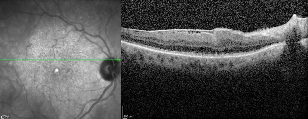
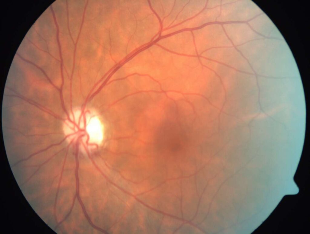
Normal fundus appearance in a healthy eye
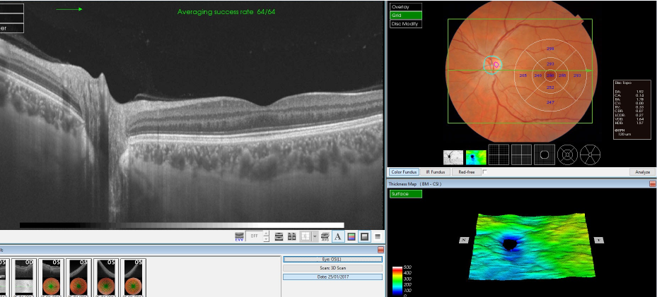
Optical coherence tomography of the macula and optic nerve in a healthy eye

Red reflex test. The examiner stands 1 m away from the patient and shines the bright light of the ophthalmoscope to cover both eyes of the patient simultaneously
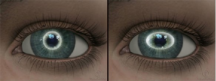
Normal corneal appearance in an eye with no astigmatism (left) and difference in the principal corneal meridians in an eye with astigmatism (right)

Ectropion (outward turning) of the right lower eyelid
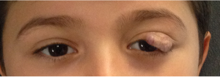
Ptosis of the left upper eyelid resulting from capillary hemangioma
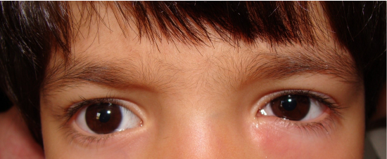
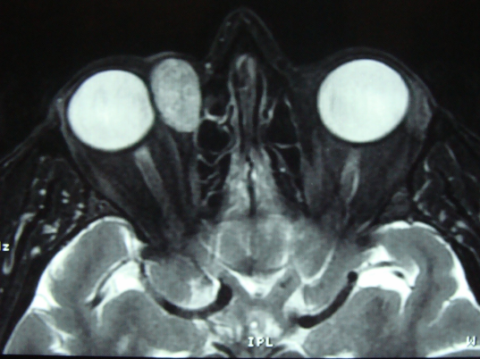
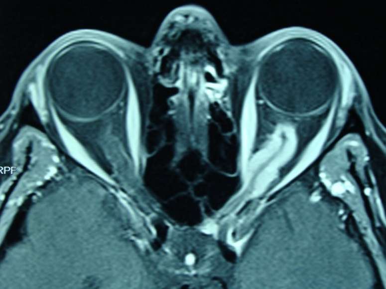
Epiphora in the left eye. This patient has chronic dacryocystitis. Magnetic resonance image of this patient is depicted in the next figure
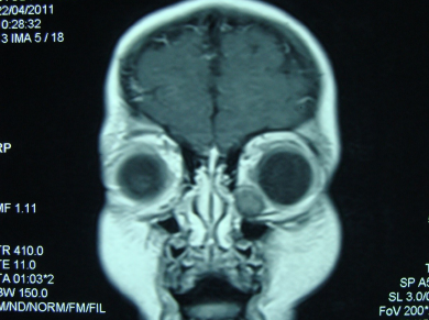
Coronal section orbital magnetic resonance imaging shows the inferonasally located dacryocystitis
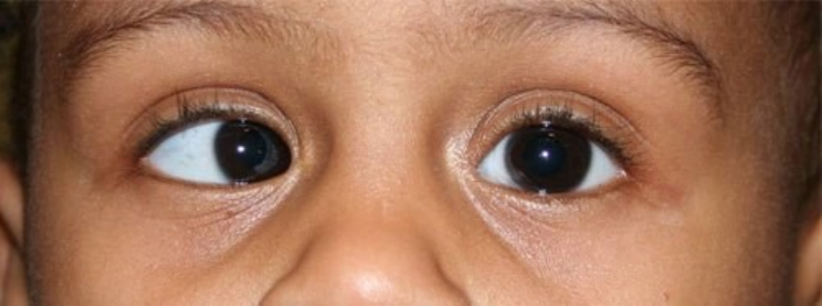
Esotropia (in-turning or inward deviation) affecting the right eye

Adenoviral conjunctivitis affecting both eyes
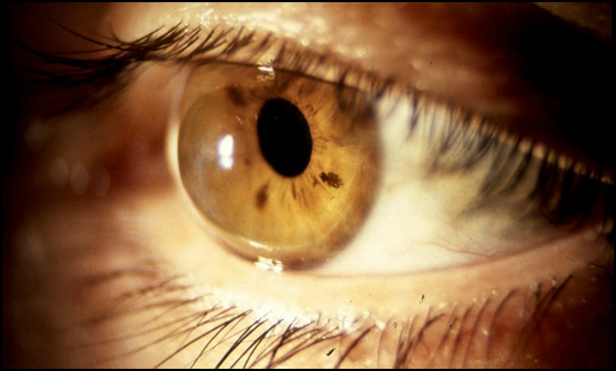
Keratoconus manifesting with steep cornea and deep anterior chamber
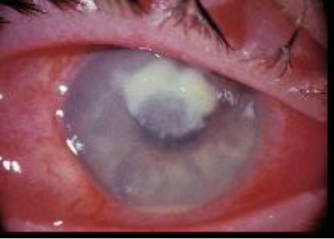
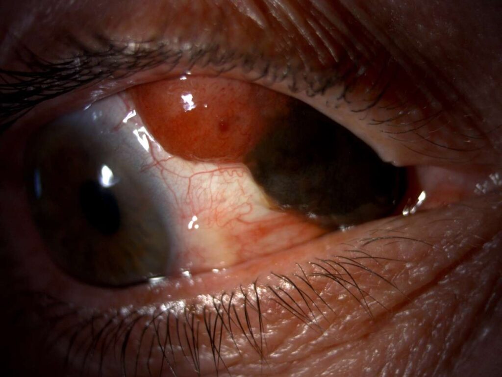
Keratitis (corneal inflammation) accompanied by hypopyon
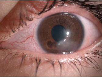
Vascularized corneal scarring in an eye with a history of keratitis
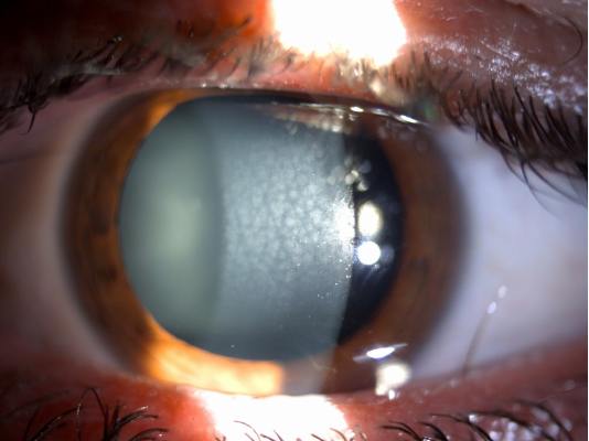
Macular type corneal stromal dystrophy
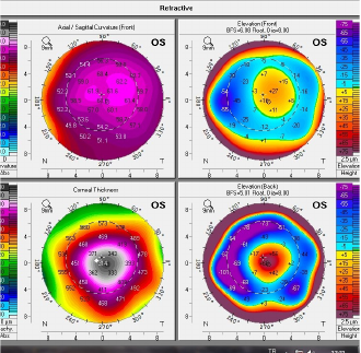
Corneal topography of an eye with keratoconus
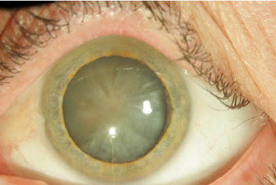
Mature cataract
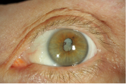
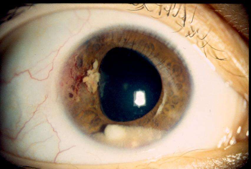
Posterior synechiae and dense cataract in a patient with choroidal melanoma after Iodine-125 plaque brachytherapy
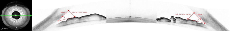
Optical coherence tomography imaging of the anterior chamber angle in an eye with open angles
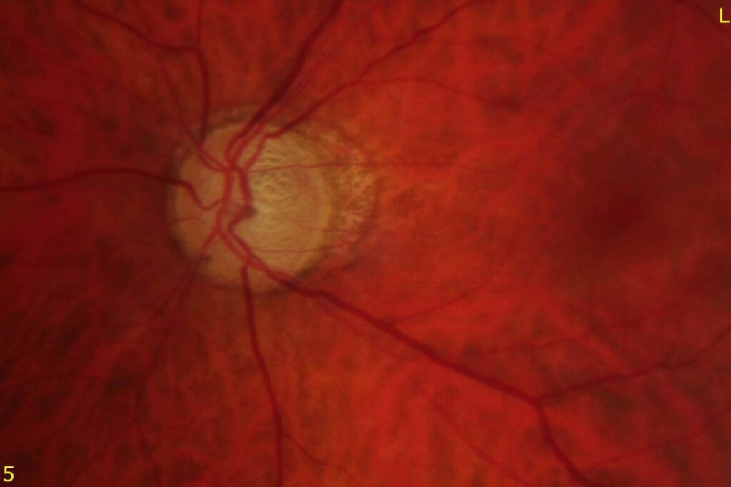
Glaucomatous optic atrophy with a marked increase in cup/disc ratio to 0.9 and peripapillary atrophy
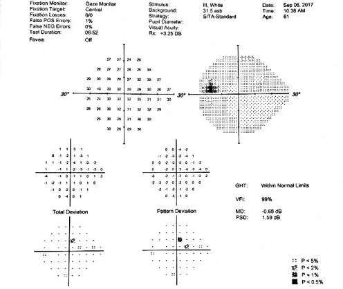
Normal visual field in a healthy eye
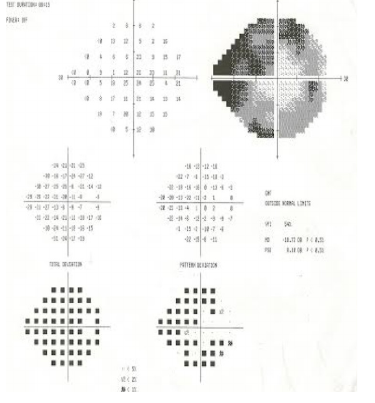
Glaucomatous visual field defect in the form of inferior and superior arcuate defects
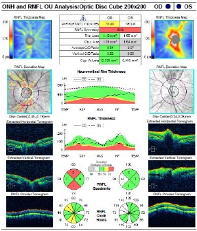
Nerve fiber layer thinning in the left eye of a patient with glaucoma. The nerve fiber layer in the right eye has normal thickness
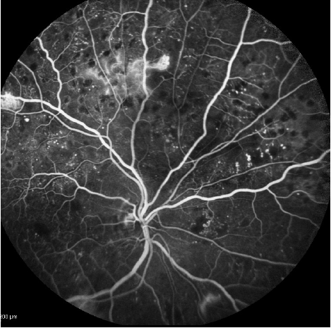
Retinal microaneurysms, hemorrhages, intraretinal microvascular abnormalities, and diabetic eye as seen on fluorescein angiography
Fundus photograph shows the thickened posterior hyaloid and epiretinal membranes in a patient with proliferative diabetic retinopathy
Massive cystoid macular edema in diabetic retinopathy as seen on optical coherence tomography
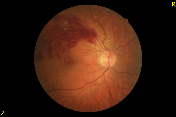
Fundus photograph shows branch retinal vein occlusion (superotemporal vein occlusion) and associated retinal hemorrhages
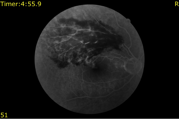
Fluorescein angiogram shows hypofluorescence due to retinal hemorrhages, retinal vessel staining, and macular edema in the late phase in an eye with branch retinal vein occlusion (depicted in the prior figure)
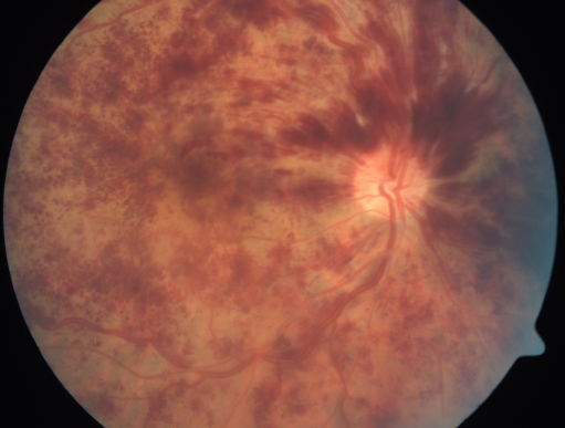
Central vein occlusion and scattered retinal hemorrhages affecting all 4 quadrants
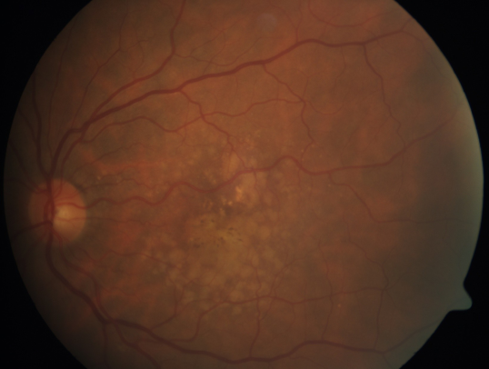
Dry type macular degeneration. Retinal pigment epithelial changes and soft drusen are noted in the macular area
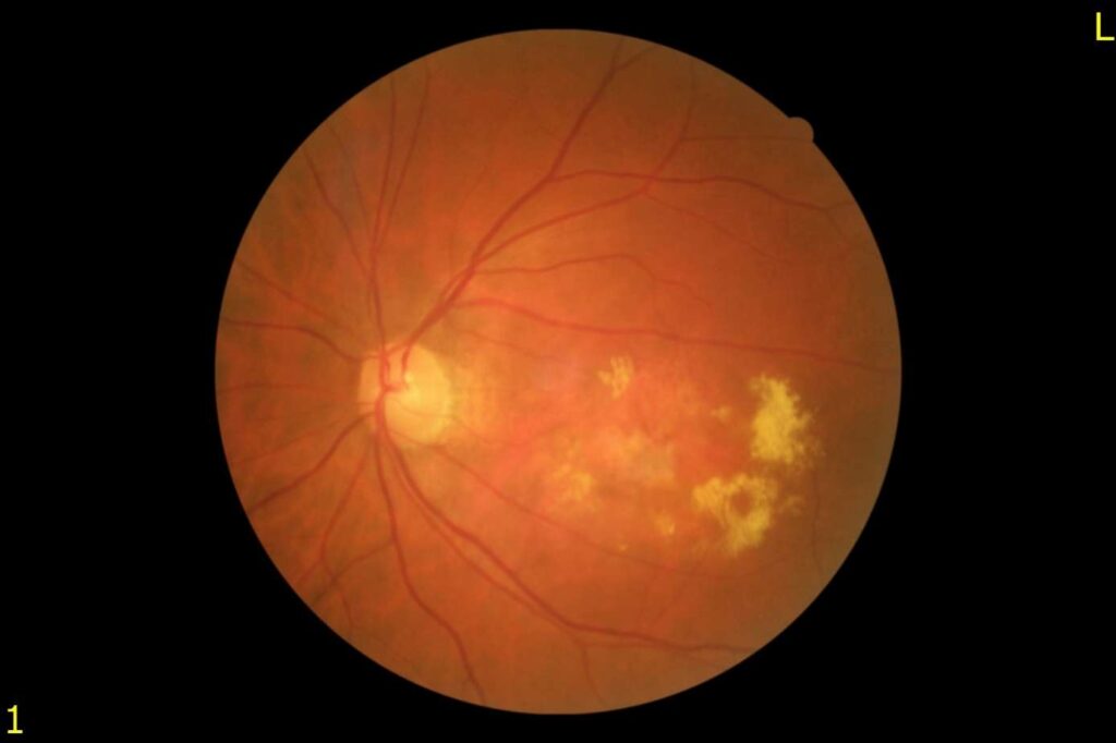
Wet type macular degeneration with choroidal neovascularization and confluent hard exudates
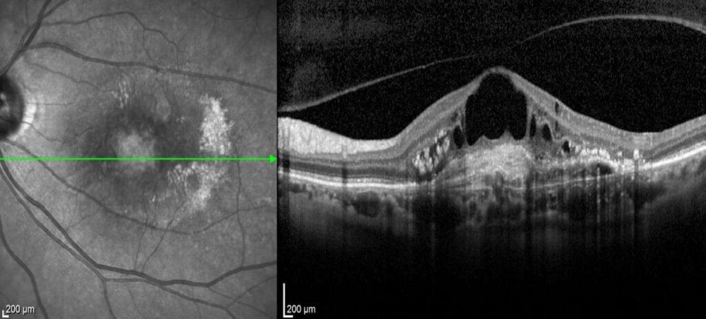
Optical coherence tomography of a patient with wet type age related macular degeneration. The detached posterior hyaloid, pigment epithelial detachment, highly reflective material under the pigment epithelial detachment (fibrin?, hemorrhage?), and massive intraretinal cystoid edema are noted
Massive submacular hemorrhage in wet type age related macular degeneration
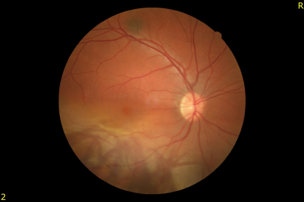
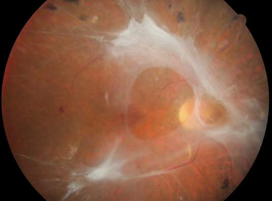
Retinal detachment affecting the inferior half of the retina
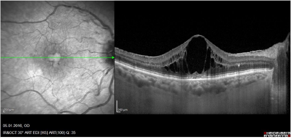
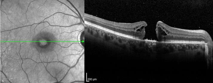
Optical coherence tomography shows macular subretinal fluid and intraretinal cysts in a patient with retinal detachment
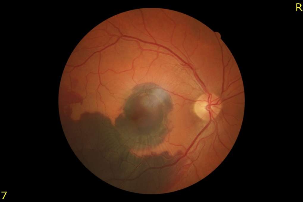
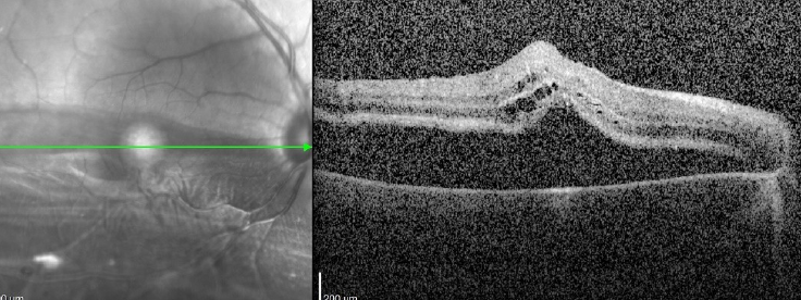
Full thickness macular hole and intraretinal edema on optical coherence tomography
Epiretinal membrane on optical coherence tomography
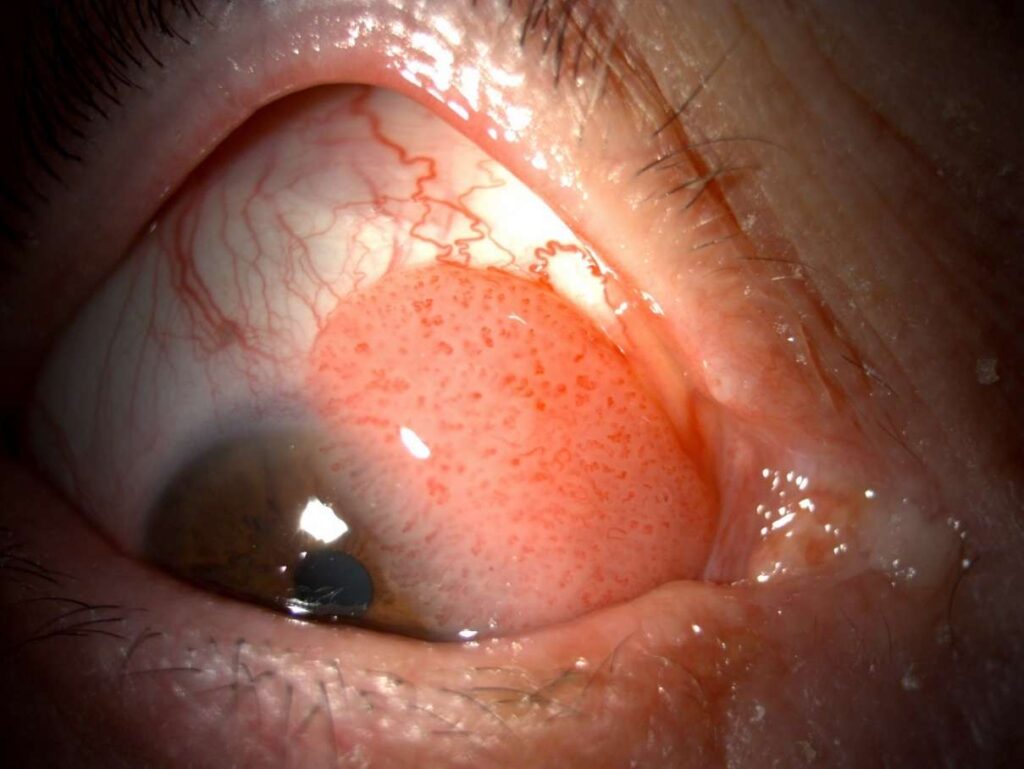
Iridocyclitis with perilimbal hyperemia and hyopyon

Ocular toxoplasmosis. A white-cream colored focus of chorioretinitis isi visible along the superotemporal vascular arcade
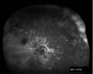
Wide-angle fluorescein angiogram shows vascular leakage, retinal hemorrhages, retinal edema, and areas of retinal non-perfusion in a patient with posterior uveitis
Upper eyelid nevus
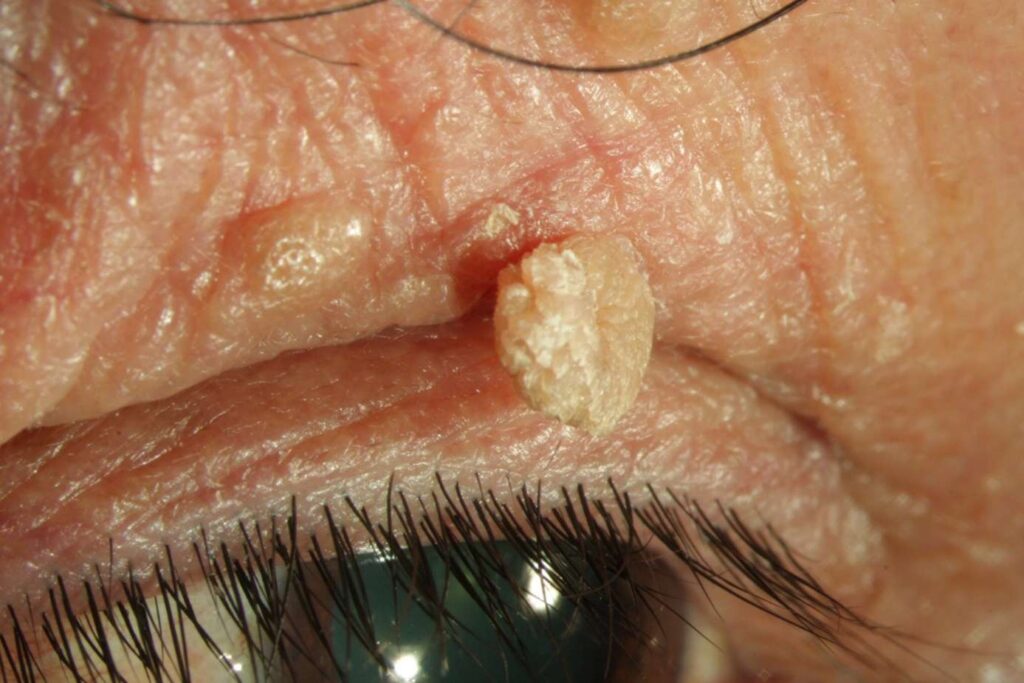
Upper eyelid papilloma

Basal cell carcinoma affecting the medial portion of the left lower eyelid. The lesion has an ulcerated appearance and there is loss of eyelashes in the medial part of the left lower eyelid
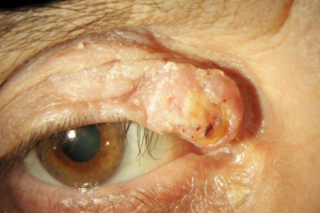
Squamous cell carcinoma affecting the right upper eyelid. The lesion has an ulcerative surface.
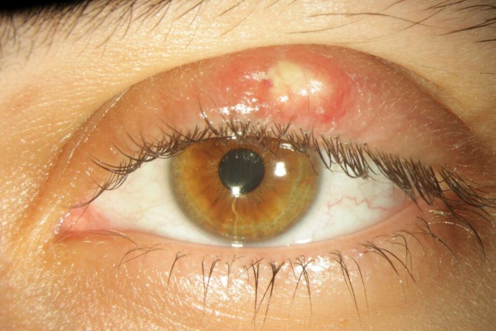
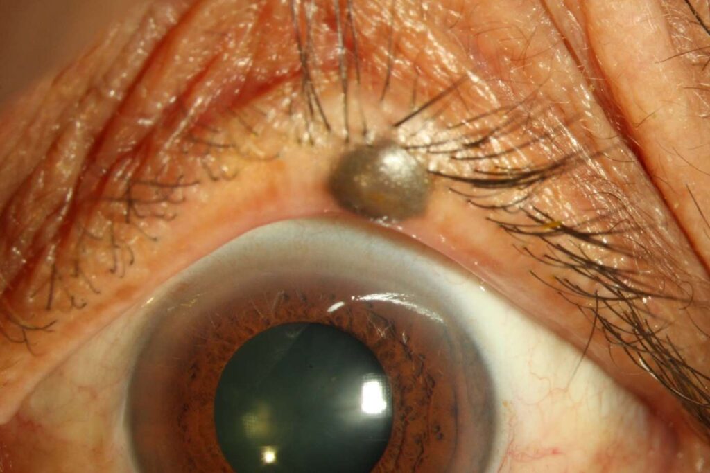
Hordeolum (stye) in the left upper eyelid
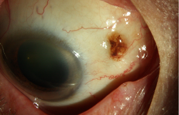
Conjunctival nevus involving the medial conjunctiva in the right eye. Intralesional cysts are seen
Squamous cell carcinoma affecting the medial and superior conjunctiva in the right eye
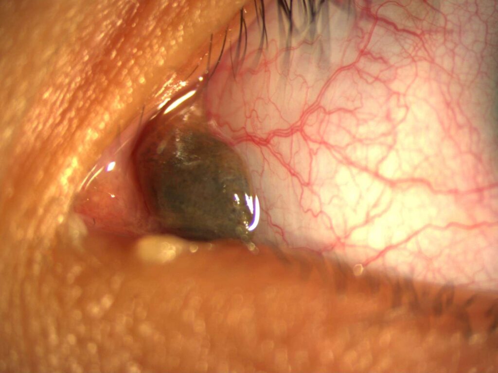
Conjunctival melanoma with pigmented and amelanotic components involving the superior fornix of the right eye
Conjunctival nevus affected the plica semilunaris of the left eye
Choroidal melanoma affecting the posterior pole and macular region of the right eye
Choroidal hemangioma along the inferotemporal vascular arcade in the left eye
Iridociliary melanoma in the inferotemporal quadrant of the left eye
Appearance of the eye depicted in the previous photograph after total tumor excision following iridocyclectomy surgery. A wide basal iridectomy is noted
An amelanotic peripapillary choroidal melanoma
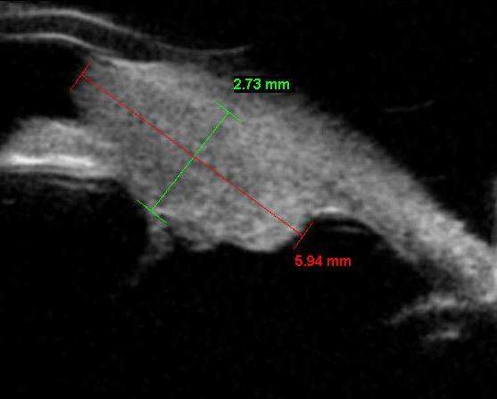
B-mode ultrasonographic appearance of choroidal melanoma demonstrating a plateau shaped tumor configuration
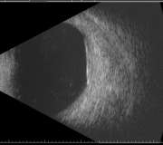
Ultrasonic bipmicroscopic appearance of ciliary body melanoma
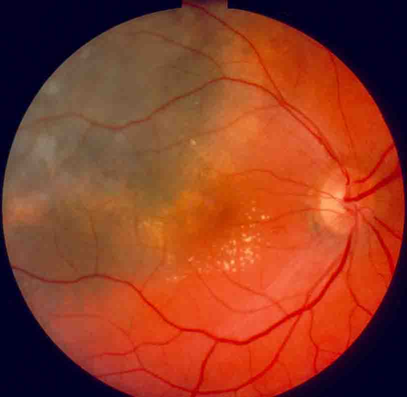
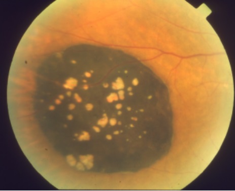
Composite fundus photograph shows congenital hypertrophy of the retinal pigment epithelium
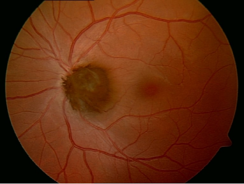
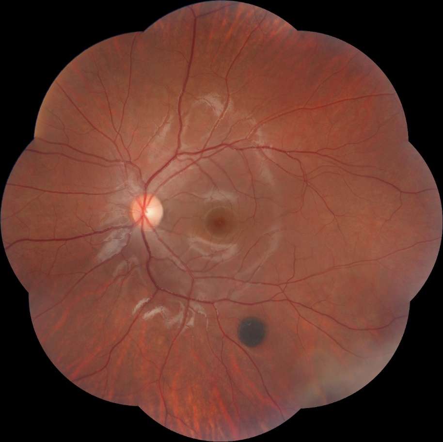
Optic nerve head melanocytoma in the left eye
Medulloepithelioma: Tumor infiltration on the iris and anterior chamber
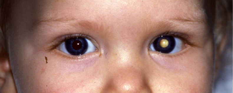
Left lekocoria. The left eye harbors retinoblastoma
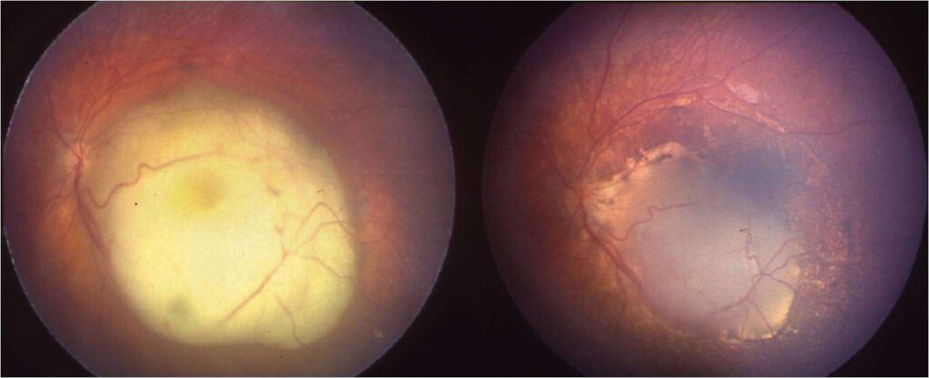
Left: Retinoblastoma affecting the macula. Right: Regression of the tumor following systemic chemotherapy
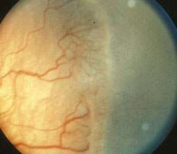
Retinopathy of prematurity (ROP) stage 2 showing the ridge formation between the vascular and more peripheral avascular retina. Advanced stages of ROP, previously called retrolental fibroplasia, may cause leukocoria and is listed in the differential diagnosis of retinoblastoma
Congenital hypertrophy of retinal pigment epithelium. Lacunae are observed in the lesion
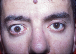
Right upper eyelid retraction in noninfiltrative thyroid ophthalmology
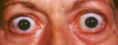
Bilateral proptosis in infiltrative thyroid ophthalmopathy
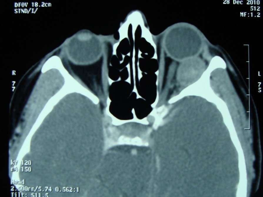
Computed tomography shows a well-circumscribed orbital tumor in the left orbit causing compression of the globe and left proptosis
Axial T2-weighted magnetic resonance image showing a well-circumscribed orbital mass affecting the right medial orbit
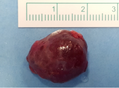
Gross photograph of orbital cavernous hemangioma after total excision via orbitotomy
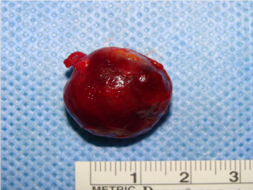
Gross photograph of orbital hemangiopericytoma after total excision via orbitotomy. Hemangiopericytoma is currently considered under the rubric of solitary fibrous tumor
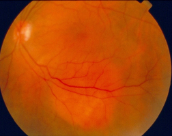
Optic atrophy originating from optic nerve sheath meningioma
Left optic nerve sheath meningioma demonstrating a tram-track appearance on axial T1-weighted contrast-enhanced magnetic resonance imaging
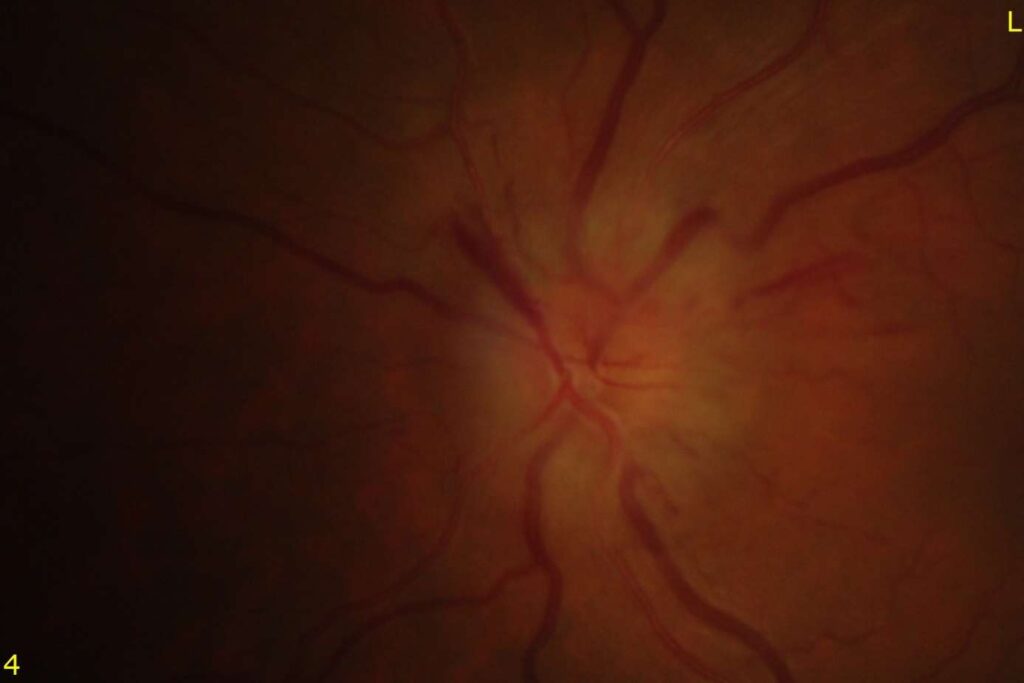
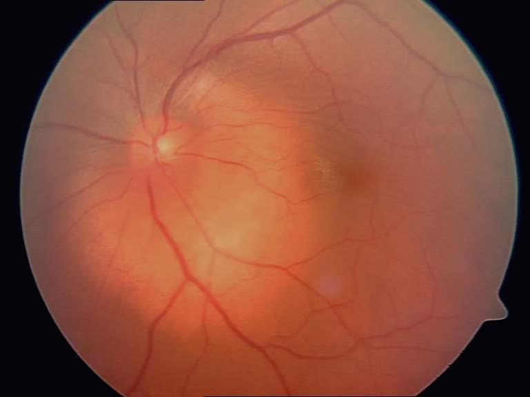
Optic nerve head edema and upper nasal splinter hemorrhage in non-arteritic ischemic optic neuropathy